
Private Skin Cancer Treatment in the UK
Non-melanoma skin cancer is highly treatable when managed by the right specialist with the right approach. For most of my patients, that approach is radiotherapy. It is the most effective, least invasive treatment available for basal cell carcinoma and squamous cell carcinoma, delivering high cure rates and cosmetic outcomes that surgery simply cannot match in many cases.
I offer rapid access to private care in London, with all treatment personally led by me from your first consultation through to discharge.
Trusted by the UK's leading medical institutions



What is Non-Melanoma Skin Cancer?
Non-melanoma skin cancer develops in the outer layers of the skin and is broadly divided into two main types: basal cell carcinoma and squamous cell carcinoma. BCC arises from the basal cells at the base of the epidermis, while SCC develops from the squamous cells in the outer layers. Both are directly linked to cumulative sun exposure over time, which is why they most commonly appear on the face, ears, scalp, neck, and hands.
While neither type carries the same metastatic risk as melanoma, they should never be left untreated. BCC can cause significant local destruction to skin, tissue, and cartilage if ignored. SCC carries a real risk of spreading to regional lymph nodes, particularly in patients with high-risk features or a compromised immune system.
The right treatment depends on the type, location, size, and growth pattern of the cancer. For most of my patients, radiotherapy is the most effective option available.
Book your consultation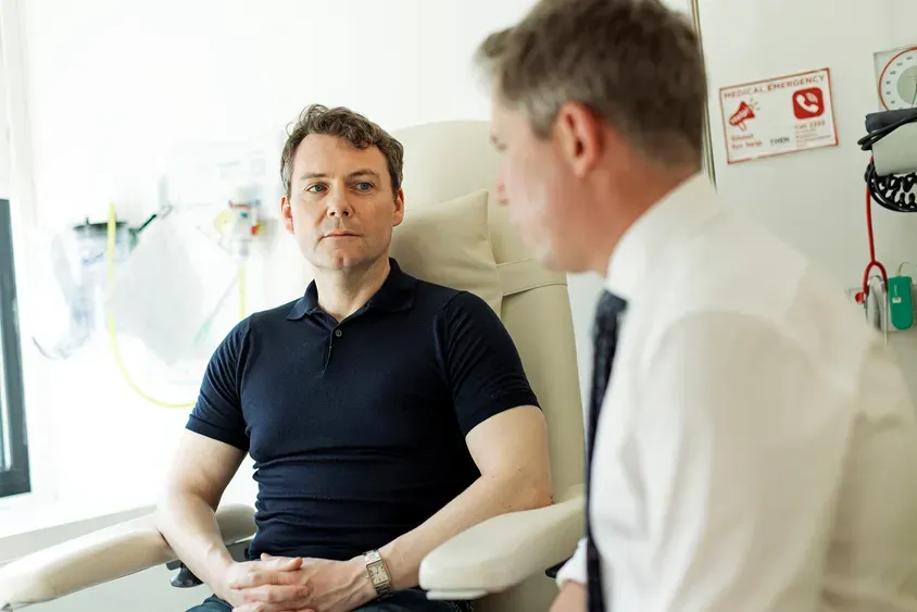
Radiotherapy vs Mohs Surgery: Which Is the Better Choice?
Both radiotherapy and Mohs micrographic surgery are highly effective treatments for non-melanoma skin cancer. For many of my patients, however, radiotherapy consistently delivers the better overall outcome when cure rate, cosmetic result, and quality of life are considered together.
| Radiotherapy | Mohs Surgery | |
|---|---|---|
| Procedure type | Non-surgical, no incision | Surgical, tissue removal required |
| Anaesthetic | Not required | Local anaesthetic required |
| Scarring | No surgical scar | Scarring dependent on reconstruction, potential tissue loss e.g. nose and ears |
| Recovery time | Some scabbing after treatment | Days to weeks depending on reconstruction |
| Sessions required | 5 to 10 short sessions | Single procedure, multiple stages |
| Cosmetic outcome at 1 year | Good to excellent in 85 to 90% of patients | Good to excellent depending on site |
| 5-year local control for BCC | 92 to 97% | 98 to 99% |
| 5-year local control for SCC | 90 to 95% | 97 to 98% |
| Best suited for | Facial lesions, elderly patients, medically inoperable, multiple lesions, patient preference | Young patients |
While Mohs surgery shows marginally higher cure rates on paper, the difference is clinically small and applies only to carefully selected high-risk cases. For the majority of my patients, particularly those with lesions on cosmetically sensitive areas of the face, radiotherapy produces outcomes that are equal or superior to surgery, without a single incision, without scarring, and without a recovery period that disrupts daily life.
The right treatment is always the one that fits the patient. But for people over 60 with cancers in a cosmetically sensitive area, radiotherapy is that treatment.
What is Radiotherapy (RT)?
Radiotherapy uses precisely targeted, carefully calculated doses of radiation to destroy cancer cells while sparing the surrounding healthy tissue.
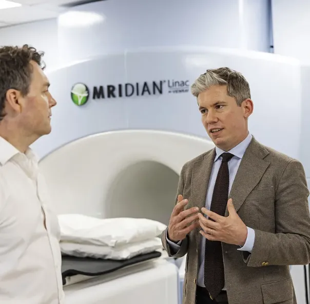
For skin cancer, external beam radiotherapy (EBRT) is one of my go-to treatments. Modern planning and delivery techniques mean I can achieve excellent cosmetic outcomes and high cure rates, particularly for BCC and SCC on the face.
Treatment is painless and delivered as an outpatient, usually over a small number of short sessions. There is no anaesthetic, no cutting and no surgical wound to care for.
Book your consultation
Basal Cell Carcinoma (BCC)
Basal cell carcinoma is the most frequently diagnosed skin cancer in the United Kingdom. Although it grows slowly and almost never spreads to other parts of the body, it will cause significant and progressive local destruction if left untreated.
Invasion of adjacent tissue, cartilage, and in some cases bone occurs over time, which is why early treatment produces both the highest cure rates and the finest cosmetic results.

How We Define Risk in BCC
Every basal cell carcinoma is classified as low risk or high risk based on both clinical and pathological features. This classification is the foundation of the treatment plan. The following features are assessed for every patient.
-
Location and size
The location of the BCC on the body and its size both contribute to the overall risk assessment. Lesions on the face, particularly around the nose, eyes, and ears, and lesions over 2 cm in diameter carry a higher risk classification.
-
Borders
Poorly defined borders indicate a more infiltrative growth pattern and raise the risk classification. Well-defined borders are a reassuring feature.
-
Primary vs recurrent
A BCC presenting for the first time carries a lower risk profile than one that has previously been treated and recurred. Recurrent lesions require more careful management.
-
Immunosuppression
Patients who are immunosuppressed, including organ transplant recipients and those on long-term immunosuppressive medications, face a higher risk of aggressive behaviour and should be assessed with particular care.
-
Site of previous radiotherapy
A BCC arising at a site that has previously been treated with radiotherapy presents additional clinical complexity and this history is always factored into the treatment recommendation.
-
Growth pattern
An infiltrative growth pattern, including morphoeic, infiltrating, or micronodular subtypes, carries a higher risk classification compared with superficial or nodular BCC. The growth pattern is identified from the biopsy result.
-
Differentiation
The presence of basosquamous differentiation raises the pathological risk classification and influences the treatment recommendation.
-
Level of invasion
Tumour invasion extending beyond subcutaneous fat indicates a more aggressive lesion requiring a more thorough treatment approach.
-
TNM stage
Lesions staged at pT2, pT3, or pT4, meaning tumours greater than 20 mm in diameter or with major bone invasion, are classified as high risk.
-
Margins
Margins that are involved by tumour, or that are closer than 1 mm, indicate a higher risk of local recurrence and must be addressed in the treatment plan.
Radiotherapy is a highly effective option across many BCC presentations. It is particularly valuable in cosmetically sensitive areas, for recurrent disease, and for patients who are not suited to surgery, delivering high cure rates while preserving the surrounding healthy tissue and appearance.
When to Offer Radiotherapy for BCC
- Lesions on the nose, ears, eyelids or lips where surgery may distort appearance
- Older or frail patients not suited to an operation
- Recurrent BCC following previous surgery
- Large or poorly defined tumours difficult to close surgically
- Patients who wish to avoid surgery
- Incomplete excision where further surgery is not feasible
When Radiotherapy Is Not Recommended for BCC
- Very young patients with long-term late-effect concerns
- Patients with Gorlin syndrome or xeroderma pigmentosum
- Previously irradiated skin that has had a full dose
- Tumours over areas of poor blood supply
- Cases where surgery offers a more reliable result with acceptable cosmesis
Squamous Cell Carcinoma (SCC)
Squamous cell carcinoma is the second most common non-melanoma skin cancer in the United Kingdom. It can progress more quickly than BCC and carries a meaningful risk of spreading to regional lymph nodes if treatment is delayed.
Unlike BCC, SCC requires prompt specialist attention. High-risk features, immunosuppression, and delayed treatment all significantly increase the chance of regional spread and a more complex management pathway.

How We Define Risk in SCC
Risk in squamous cell carcinoma is assessed not only to guide the initial treatment decision but also to determine the follow-up protocol after treatment is complete. Low-risk, high-risk, and very high-risk SCC each carry a different surveillance schedule, and getting that classification right from the outset is essential to protecting the patient long term.
-
Size
Tumours greater than 2 cm carry a significantly higher risk of recurrence and regional spread. Size is one of the strongest independent predictors of metastatic risk in SCC.
-
Depth
Lesions deeper than 6 mm, or those invading beyond subcutaneous fat, are classified as high risk regardless of other features.
-
Location
SCC arising on the ear, lip, temple, or non-sun-exposed sites such as the genitalia or perianal region carries a substantially higher risk of nodal spread than lesions on the trunk or limbs.
-
Immunosuppression
Organ transplant recipients and patients on long-term immunosuppressive therapy face a significantly higher risk of aggressive SCC with greater metastatic potential and reduced treatment response.
-
Recurrent disease
A recurrent SCC carries a much higher risk profile than a primary lesion. Prior treatment failure indicates a more aggressive tumour biology and demands a more thorough management approach.
-
Differentiation
Poorly differentiated or undifferentiated SCC carries a substantially worse prognosis than well-differentiated tumours. Grade is one of the most important pathological risk factors assessed at biopsy.
-
Perineural invasion
The presence of perineural invasion, particularly involving a named nerve, when multifocal, or extending deeper than 0.1 mm below the dermis, significantly increases the risk of recurrence and is a strong indication for adjuvant radiotherapy.
-
Lymphovascular invasion
Evidence of lymphovascular invasion on histology indicates a higher likelihood of regional nodal spread and escalates the overall risk classification.
-
TNM stage
Tumours staged at T2 or above, including those greater than 2 cm, those with deep invasion, or those with bone involvement, are classified as high risk and managed with a more aggressive treatment and surveillance protocol.
-
Margins
Involved or close margins following surgical excision indicate incomplete removal and are a direct indication for adjuvant radiotherapy where further surgery is not possible.
Radiotherapy plays a central role in SCC management across multiple stages of the treatment pathway. It can be used as the primary treatment when surgery is not appropriate, and as an adjuvant treatment following surgery to significantly reduce the risk of recurrence. For patients with perineural invasion, incomplete excision margins, or recurrent disease, adjuvant radiotherapy is frequently the most important intervention in the entire treatment plan.
When to Offer Radiotherapy for SCC
- Surgery is not clinically feasible for the patient
- Surgery would cause unacceptable functional or cosmetic outcome
- SCC incompletely excised and further surgery not possible
- Perineural invasion involving a named nerve or below dermis
- Recurrent disease following previous treatment attempt
- Immunocompromised patients presenting with high-risk features
- Excision margins are clear but closer than 1 mm
When Radiotherapy Is Not Recommended for SCC
- Young patients with significant long-term late effect concerns
- Tumours located in areas of poor vascular supply
- Bone, cartilage, or tendon involvement without mutilating alternative
- Patients diagnosed with Gorlin syndrome or xeroderma pigmentosum
- Cases where long-term cosmetic deterioration outweighs surgical risk
Radiotherapy Results: Before and After Treatment
Every image below represents a real patient treated personally by Dr Wilson using external beam radiotherapy. The results speak for themselves. Outstanding cancer control, no surgical scarring, and cosmetic outcomes that consistently meet or exceed what surgery can achieve in the same locations.
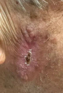
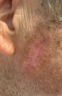
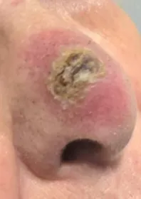
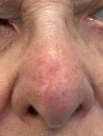
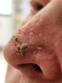
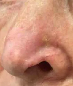
Dr Wilson offers same or next day appointments across four London locations. You will speak directly with a consultant oncologist who will assess your situation carefully, explain your options in plain English and design a treatment plan built around you as an individual.
From First Consultation to Complete Recovery
Every stage of your care is led personally by Dr Wilson. From your first appointment through to your final follow-up review, you will always be seen by the same consultant who knows your case in full. There are no unexpected handovers, no unexplained gaps, and no moments where you are left without a clear point of contact.
Initial Consultation
A private consultation with Dr Wilson, typically available within 48 hours. He will examine the lesion, review your history, and give you a clear picture of what comes next.
Confirmation of Treatment Plan
Your treatment plan is confirmed personally by Dr Wilson, with all findings reviewed and discussed before your first session begins.
Treatment and Delivery
Delivered at The Cromwell Hospital or one of our London locations. Sessions last only a few minutes and most patients return to normal daily activity the same day.
Aftercare and Surveillance
Your follow-up schedule is tailored to your risk level, with Dr Wilson as your direct point of contact from post-treatment review through to long-term surveillance.
What People Are Saying
The most honest measure of care is the experience of those who have received it. These reviews come from real patients who trusted Dr Wilson at one of the most difficult times in their lives, and wanted to share what that experience meant to them.


Frequently asked questions
-
What is the most effective treatment for basal cell carcinoma?
The most effective treatment for basal cell carcinoma depends on the specific characteristics of your cancer. For many patients, radiotherapy offers cure rates comparable to surgery with excellent cosmetic outcomes, particularly for facial lesions. Your treatment will be personalised based on the location, size, and type of your BCC.
-
How many radiation treatments for basal cell carcinoma will I need?
Most patients receive 5-10 radiation treatments over 1-2 weeks. The exact number depends on factors including the size and location of your tumour, your general health, and the specific radiotherapy technique used. I'll create a personalised treatment plan that balances effective cancer control with minimal side effects.
-
What is the best treatment for squamous cell carcinoma?
The best treatment for squamous cell carcinoma is determined by several factors, including the tumour's size, location, depth, and aggressiveness. While surgery is common, radiotherapy offers excellent outcomes for many patients, particularly for facial lesions where cosmetic results are important. For some cases, a combination of treatments may provide optimal results.
-
Is radiotherapy as effective as surgery for skin cancer?
For properly selected cases of basal cell carcinoma and cutaneous squamous cell carcinoma, radiotherapy offers cure rates comparable to surgery. It's particularly valuable for tumours in cosmetically sensitive areas or locations where surgery might compromise function. The non-invasive nature of radiotherapy also makes it an excellent option for patients who wish to avoid surgical procedures.
-
What happens after treatment for non-melanoma skin cancer?
After completing treatment, you'll have regular follow-up appointments to monitor healing and ensure the cancer remains controlled. The treated area will gradually heal over 4-6 weeks. Long-term, you'll need to be vigilant about sun protection, as treated skin is more sensitive to UV radiation. I'll provide comprehensive guidance on post-treatment care and monitoring.
-
Why choose Dr James Wilson for non-melanoma skin cancer treatment?
As a leading Clinical Oncologist specialising in non-melanoma skin cancer treatment, I offer:
- Expertise in cutting-edge radiotherapy techniques
- Access to innovative treatments including Rhenium-SCT
- A comprehensive, personalised approach to your care
- Focus on both cancer control and cosmetic outcomes
- Rapid access to consultations, tests and treatments
- Continuity of care throughout your entire treatment journey
At every appointment, you'll see me personally, not a doctor-in-training. I ensure all your questions are answered and share my treatment letters and reports with you on the same day as your appointment.
Straight From the Clinic
Dr Wilson shares clear, expert insight on skin cancer, radiotherapy and what to expect from treatment.
Subscribe to the channel and get notified every time a new video is published.